
What are the symptoms of Diseased Aortic heart valve?
If heart valves begin to fail, the heart beats harder to compensate for the reduced blood flow. As the heart beats harder, symptoms of valve disease may occur:
- You may have Increased shortness of breath Heart palpitations and feeling of skipped heartbeats
- You will have Chest pain or discomfort Swelling of the ankles, feet or abdomen (edema) and Rapid weight gain (3 pounds in one day is possible)
- Increasing fatigue and weakness or dizziness
Symptoms can occur quickly if the onset of valve disease is severe and sudden. For some people, symptoms may occur gradually and may not be noticeable.

How to diagnose diseased aortic heart valve ? What are the different ways to diagnose it
Aortic Heart Valve diseases are diagnosed after your doctor evaluates your symptoms and the results of your physical exam and diagnostic tests. The physical exam may reveal fluid in the lungs, an enlarged heart or a heart murmur, the sound made by blood moving through a stenotic or a leaky valve.
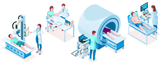
5 Ways to diagnose diseased Aortic heart valve
Diagnostic tests help your doctor identify the location, type and extent of your valve disease and may include: During the exam, your physician will listen to your heart. One indication of valve disease is the presence of a murmur, an abnormal sound caused by turbulent blood flow across a valve. A heart murmur does not always indicate a heart valve problem. However, most abnormal heart valves cause a murmur.
- Echocardiogram- An echocardiogram uses high frequency sound waves to determine how the parts of the heart are working. This allows the physician to determine valve leakage or to measure the opening of a stenotic valve.
- Transesophageal echocardiogram- A transesophageal echocardiogram (TEE) also uses sound waves to look at the heart and measure a valve opening or to determine leakage. This test differs from a standard echocardiogram because a probe is inserted into the esophagus. In this procedure, the probes its directly behind the heart, allowing certain parts of the heart to be seen more clearly than with the standard test.
- Cardiac angiogram or cardiac catheterization- A cardiac angiogram or cardiac catheterization looks at blood flow to the heart, helping detect the area and extent of any blockage or narrowing of the arteries. During the procedure, a thin tube is inserted into the femoral artery (located in the groin) and passed into the heart. Contrast dye is injected into the tube and X-rays are taken, allowing blood vessels and valves to be seen clearly on the X-ray.
- Cardiac magnetic resonance imaging- cardiac magnetic resonance imaging uses a magnetic field and radio waves to create detailed images of the heart chambers, blood vessels and valves.
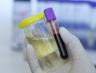
5. Blood and urine test- Blood and urine tests to check for kidney problems, blood sugar levels, and the blood’s ability to clot.